![[55Fe,57Co]](xr100cr_a.gif)
Figure 7. 55Fe, 57Co, and Test Pulser.
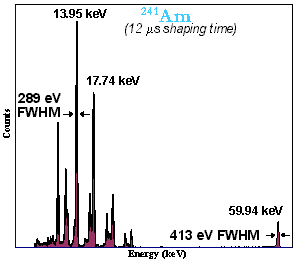
Figure 9. 241Am Spectrum.
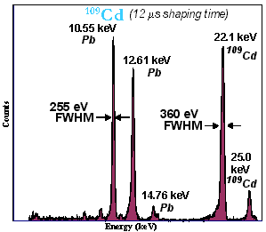
Figure 10. Lead (Pb) Fluorescence from 109Cd.
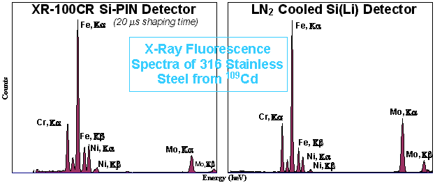
Figure 11. Amptek XR-100CR Si-PIN Detector Compared with Si(Li) Detector.

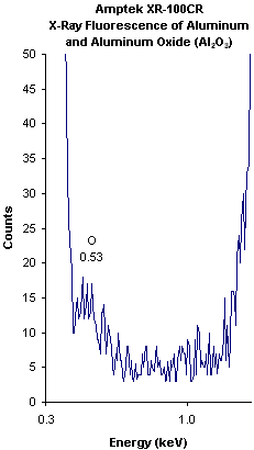
Low Energy Performance of the XR-100CR
![[55Fe,57Co]](xr100cr_a.gif) Figure 7. 55Fe, 57Co, and Test Pulser. |
 Figure 9. 241Am Spectrum. |
 Figure 10. Lead (Pb) Fluorescence from 109Cd. | |
 Figure 11. Amptek XR-100CR Si-PIN Detector Compared with Si(Li) Detector. | |
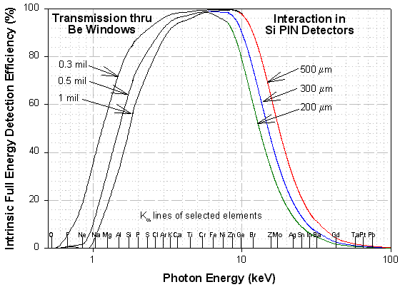 Figure 2. |
Figure 2 (linear) shows the intrinsic full energy detection efficiency for the XR-100CR detectors. This efficiency corresponds to the probability that an X-ray will enter the front of the detector and deposit all of its energy inside the detector via the photoelectric effect.
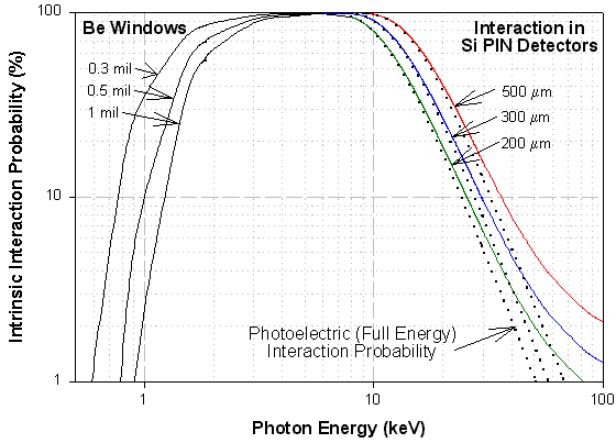 Figure 3. |
Figure 3 (log) shows the probability of a photon undergoing any interaction, along with the probability of a photoelectric interaction which results in total energy deposition. As shown, the photoelectric effect is dominant at low energies but at higher energies above about 40 keV the photons undergo Compton scattering, depositing less than the full energy in the detector.
Both figures above combine the effects of transmission through the Beryllium window (including the protective coating), and interaction in the silicon detector. The low energy portion of the curves is dominated by the thickness of the Beryllium window, while the high energy portion is dominated by the thickness of the active depth of the Si detector. Depending on the window chosen, 90% of the incident photons reach the detector at energies ranging from 2 to 3 keV. Depending on the detector chosen, 90% of the photons are detected at energies up to 9 to 12 keV.
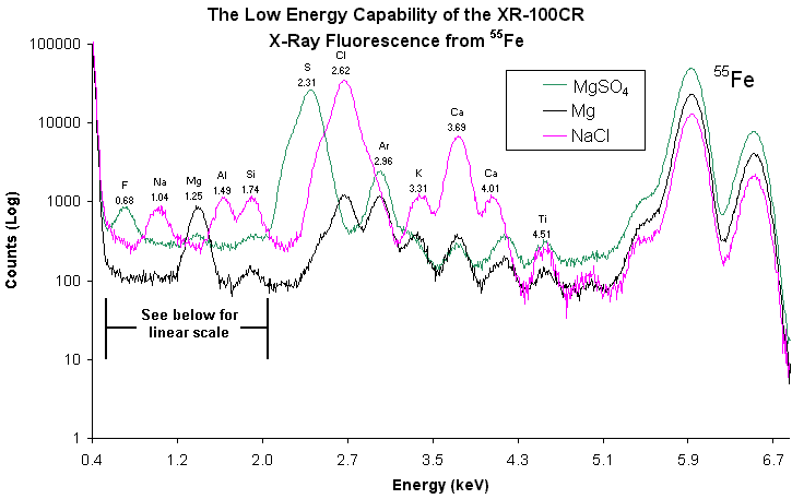
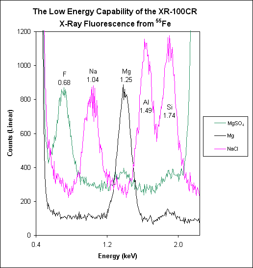
The figures above show the performance of the XR-100CR down to Fluorine (0.68 keV). The spectra were taken with a standard 7mm2 detector, a 0.3 mil (8µm) Berylium window, an internal Tungsten (W) collimator, and a 20µs shaper. The samples were Epsom Salt (MgSO4), Magnesium (Mg), and common salt (NaCl) irradiated by an 55Fe source.
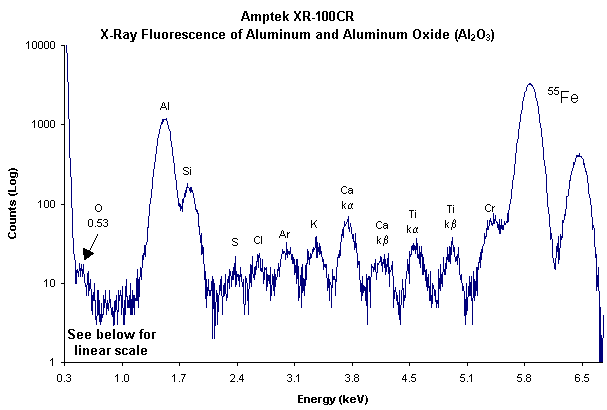
Amptek X-Ray Chart (K and L emission lines)
Revised February 6, 2001