
Medical X-Ray Tube Spectra
for Mammography and Radiology
X-Ray Tube Monitor for Mammography Machines
![[molybdenum spectrum]](medical2.gif)
X-Ray Tube Monitor for Radiology Machines
![[W Spectrum]](medical1.gif)
Spectra Courtesy of
Dr. Andrew Karellas Ph.D
University of Massachusetts Medical School
Worcester, MA. 01655 USA
|
Direct Measurement SpectraEnd Point Energy (kVp)See what the patient gets
- NO Compton Spectra
- NO Corrections
- NO Calculations
Self-Calibrating SystemLook straight at the X-Ray tube and record simultaneously both the spectrum and the peak potential (kVp)The technology that went to Mars on the Pathfinder Mission is now available to Radiology!A must detector for every Radiology DepartmentFor Quality Assurance in Radiographic and Fluoroscopic Systems- No Liquid Nitrogen
|
Design Objective
This detector system was designed with the objective of simultaneously measuring the X-Ray tube peak potential (kVp), and
to characterize the mammographic X-Ray tube spectrum.
Significance of the Measurement
- Both the tube spectrum and the peak potential (kVp) are important parameters affecting the image quality in film-screen
and digital mammography.
- Automatic selection of proper target/filter combination in modern mammography systems maybe affected by improper kVp.
- In conventional devices, the user depends on central laboratory calibration and has no easy way to calibrate the instrument
during use.
Complete System Includes:
|
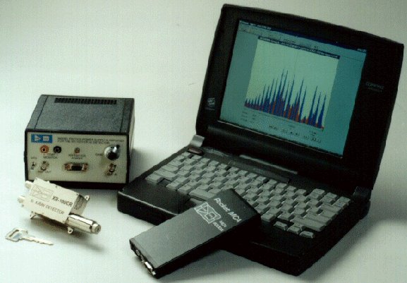
XR-100T-CZT system shown
with Amptek MCA8000A Multichannel Analyzer
and a notebook computer
|
All Solid State Design - - - No Liquid Nitrogen!!!
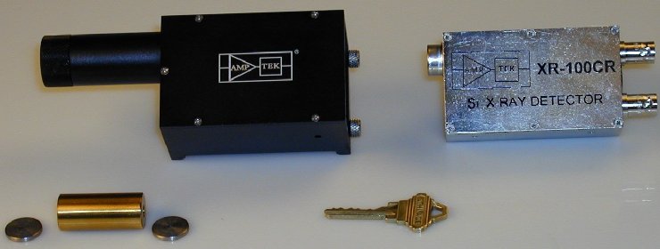
System Description
The XR-100T-CZT is a high performance X-Ray and Gamma Ray detector mounted on a thermo-electric
cooler (Peltier type) together with the input FET to the preamplifier. Monitored by an
integrated circuit, these components are kept at -30° C and are enclosed in a hermetic package with
a vacuum tight, light tight Beryllium window. Power and signal processing to the detector is
provided by the PX2T-CZT in order to ensure quick, stable operation in less than one minute from
power turn-on. The output pulse produced by the PX2T-CZT connects directly to the input of the
Multichannel Analyzer MCA8000A "Pocket MCA."
Collimator Kit
Amptek has developed a Collimator Kit to collimate the primary X-ray beam. This system is comprised
of a special Detector/Preamp box (TJ BOX) which slides inside a Collimator Housing. The Collimator Housing can accommodate up to two
Tungsten collimator disks that are placed inside a bayonet holder in front of the detector. By selecting the appropriate
Tungsten collimator disks, the user can reduce the incoming X-ray flux and allow the detector and electronics to process the
X-ray spectrum. Seven different Tungsten collimator disks are provided with different size holes (ranging from 25 µm
to 2,000 µm hole) in order to allow for a wide range of applications. The Collimator Housing is made out of brass in order to shield the detector and electronics from the primary X-ray source. Additionally, the bottom of the Collimator Housing incorporates a screw type fitting to allow for a standard tripod mount.
References
- Matsumoto, Massao, et al. Direct measurement of mammographic x-ray spectra using CdZnTe detector, Medical Physics 27 (7), July 2000. p. 1490.
- Vedantham, Srinivasan, et al. Mammographic imaging with a small CCD-based digital casette: Physical characteristics of a clinical system, Medical Physics 27 (8), August 2000.
- Vedantham, Srinivasan, et al. Full breast digital mammography with an amorphous silicon-based flat panel detector: Physical characteristics of a clinical prototype, Medical Physics 27 (3), March 2000.
Medical X-Ray Detector Specifications and description in PDF format (418k)
XR100T-CZT | MCA8000A "Pocket MCA" | Home Page | Products | Price List | Company Profile | Press Release
Revised February 7, 2001